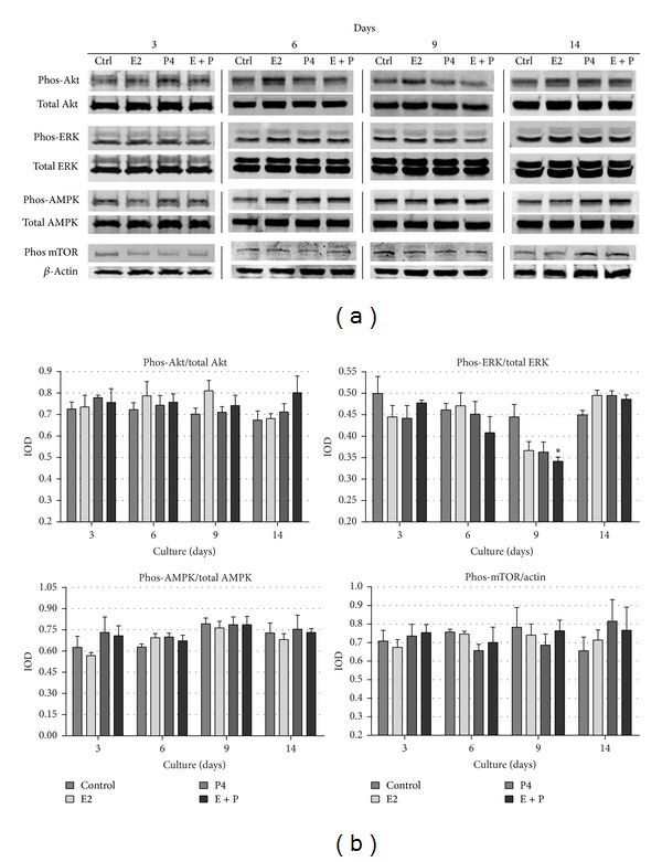
Figure 6.

(a) Western blot analysis of the levels of chosen kinases (Akt, ERK, AMPK, and mTOR) involved in autophagy regulation, detected in BME-UV1 cells cultured on Matrigel for 3, 6, 9, and 14 days in differentiation medium (control), enriched with 17β-estradiol (E2, 1 nM), progesterone (P4, 5 ng/mL), or both (E + P): expression of β-actin was used as a loading control; (b) graphs represent the obtained results of densitometric analysis, in which IOD of each band was measured, and the IOD values for phosphorylated forms of kinases were normalized to the respective IOD of the total forms, with an exception of phos-mTOR, which was normalized to IOD of β-actin; results are presented as means ± SEM from at least three separate experiments; *statistically significant difference (P < 0.05) in comparison with control conditions.
