
Figure 7.
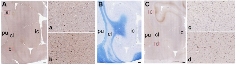
Low-magnification images of the CBPs in the human claustrum. (A) CR-immunoreactive neurons; a and b are enlargements of the corresponding red rectangles in A; (B) Reference image stained with Luxol Fast Blue; (C) PV-immunoreactive neurons; c and d are enlargements of the corresponding red rectangles in C; pu; putamen; cl: claustrum; ic: insular cortex. Scale bars: A, B, C = 1 mm; a, b, c, d, = 100 µm.