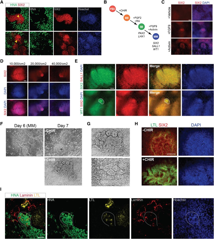
Figure 5.
FGF-9 and activin differentiate PAX2+LHX1+ cells into cells expressing markers of CM. (A) Whole-mount immunohistochemistry for anti–human nuclear antigen (HNA) and SIX2 in chimeric kidney explant cultures. Dissociated hESC-derived IM cells on day 9 (n=3) of differentiation were mixed with dissociated E12.5 mouse embryonic kidneys in single cell suspension, reaggregated by centrifugation, and cultured for 5 days in kidney explant culture. Arrowhead, HNA+ cells present within clusters of mouse Six2+ cells. Scale bar, 50 μm. (B) Diagram showing the stepwise differentiation of hESCs into metanephric CM. (C) Immunostaining for SIX2 in hESCs treated with FGF2+RA (ChFR) for 3 days then either 100 ng/ml FGF-9, 10 ng/ml activin, or vehicle for 3 days, day 6. Scale bar, 100 μm. (D) Immunostaining for SIX2 in hESCs plated at different densities and treated with ChFR for 3 days then 100 ng/ml FGF-9+10 ng/ml activin for 3 days, day 6. Scale bar, 100 μm. (E) Immunostaining for SIX2, SALL1, and WT1 in hESCs treated with ChFR for 3 days then 100 ng/ml FGF-9+10 ng/ml activin for 3 days, day 6. Dashed line encompasses the population of cells that stain positive for WT1. Scale bar, 100 μm. (F) Brightfield microscopy of hESCs treated with ChFR for 3 days, 100 ng/ml FGF-9+10 ng/ml activin for 3 days, then either 5 μM CHIR (+CHIR) or FGF-9+activin (−CHIR) for 24 hours. Scale bar, 100 μm. (G) Higher magnification of tubular epithelial-like structures seen in SIX2+ cells treated on day 6 with 5 μM CHIR for 24 hours, day 7. Scale bar, 100 μm. (H) Immunostaining (day 8) for SIX2 and LTL in hESCs treated with ChFR for 3 days, 100 ng/ml FGF-9+10 ng/ml activin for 3 days, then either 5 μM CHIR (+CHIR) or FGF9+activin (−CHIR) for 24 hours. Scale bar, 100 μm. (I) Whole-mount immunohistochemistry for anti–human nuclear antigen (HNA), laminin, and LTL in chimeric kidney explant cultures. Dissociated hESC-derived SIX2+ cells on day 6 (n=10) of differentiation were mixed with dissociated E12.5 mouse embryonic kidneys in single cell suspension, reaggregated by centrifugation, and cultured for 3 days in kidney explant culture. Dashed line encompasses an organizing cluster of HNA+ cells, which express laminin and LTL. Scale bar, 50 μm. DAPI, 4′,6-diamidino-2-phenylindole; ME, mesendoderm; MM, metanephric cap mesenchyme; PSC, pluripotent stem cell.

