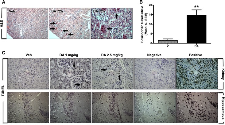
Figure 3.
Acute DA administration induced cell death and renal histopathology. (A and B) Renal histopathology was examined by hematoxylin and eosin (H&E). (A) Note the intense cytoplasmic eosinophilia of proximal tubule cells, which is indicative of cell death at 72 hours after 2.5 mg/kg DA (arrows in center panel); it was not present in vehicle kidneys (Veh; left panel). Higher-magnification view of the renal cortex depicts eosinophilic cells within the luminal space, which is indicative of tubule cell desquamation after injury (arrows in right panel). (B) Quantification of eosinophilic tubules in vehicle- and DA-treated kidneys (n=3, 8 fields per n, **P<0.01, t test). V, vehicle. (C) Apoptosis was assessed by TUNEL staining in kidney and brain sections from vehicle- and DA-treated animals. Note TUNEL-positive renal tubule cells after exposure to 1.0 (arrow) and 2.5 mg/kg DA (arrows). No TUNEL-positive cells were detected in the CA3 region of the hippocampus after DA. Negative controls were processed without terminal deoxynucleotidyl transferase enzyme, and positive controls were treated with DNase I at the beginning of the protocol. Veh, vehicle.

