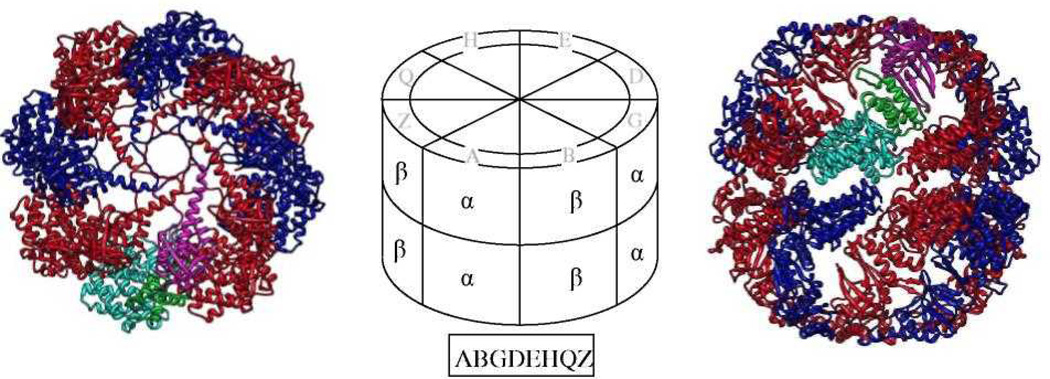
Figure 1.
Our structural model is based on the known structure of the thermosome (PDB file 1A6D), which has α (blue) and β (red) subunits that alternate around each of the two rings. The structure is viewed from the top (left) and side (right). The three domains of one of the α subunits in the top ring are colored cyan, green & magenta for the equatorial, middle & apical domains, respectively. The central schematic shows how to map the 8 subunits - A, B, G, D, E, H, Q & Z - of TRiC in a proposed arrangement (boxed) onto the top ring of the thermosome. This arrangement is brought as an example only, and is not part of our suggested set of 72.

