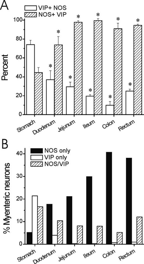
Figure 6.
Distribution and colocalization of NOS and VIP in myenteric neurons along the GI tract from rhesus monkey. A: Percentage of VIP neurons also expressing NOS (hatched bars) is higher distal to the duodenum. The percentage of NOS neurons also expressing VIP (white bars) is lower distal to the stomach. *P < 0.05 vs. stomach, ANOVA with post-hoc Bonferroni. B: Absolute percentage of neurons was calculated based on percentages determined with HuC/D colabeling (Fig. 2). There is a significant increase in the abundance of NOS-only myenteric neurons in the distal GI tract.

