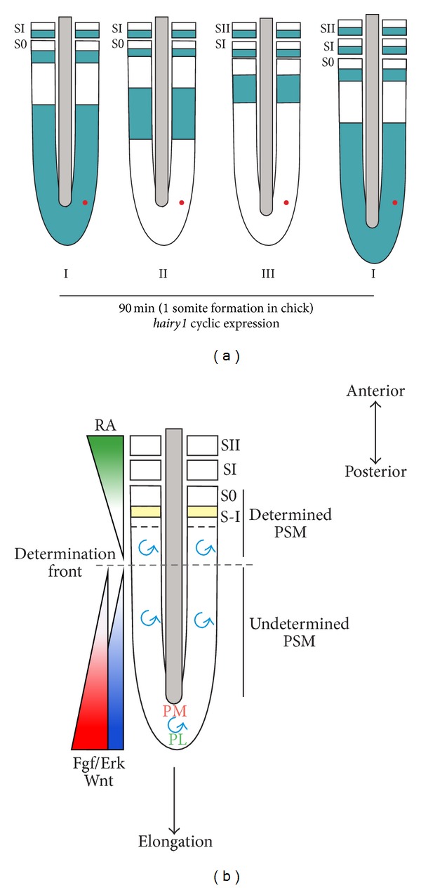
Figure 2.

The molecular clock and wavefront. (a) Representation of the segmentation clock, visualized as distinct phases of expression (I, II, and III) of the chick oscillating gene hairy1 [32]. Hairy1 mRNA transcriptional oscillations are propagated as posterior-to-anterior kinematic waves that sweep the PSM and culminate with the formation of a new pair of somites (SI) reiterated every 90 min in the chick. During each cycle, individual cells (represented as a red dot in the PSM) periodically turn on and off gene expression. (b) Integration of the signalling activities in the PSM regulating somite formation. Molecular clock oscillations (blue spiral) take place in the somitic precursor cells in the tail-bud region and along the entire PSM. Opposing gradients of Fgf (red), Wnt (purple), and retinoic acid (RA, green) position the determination front (dashed line) in the PSM [33–36]. High Fgf levels protect PSM posterior cells from precocious differentiation by inhibiting raldh2 expression, thus maintaining it in an undifferentiated state (undetermined PSM). The PSM tissue above the determination front is considered to be determined and contains three to four presumptive somites [33]. Confrontation between the molecular clock oscillations and the determination front is required to define segment formation by inducing mesp2 in the anterior PSM (yellow) [37, 38]. High Fgf/Wnt levels in the posterior PSM repress mesp2 expression [39, 40], which is activated only when Fgf/Wnt levels drop below a threshold. S0 represents the forming somite and SI and SII represent the two most recent formed somites. PM and PL represent prospective medial and lateral PSM, respectively.
