
Figure 1.
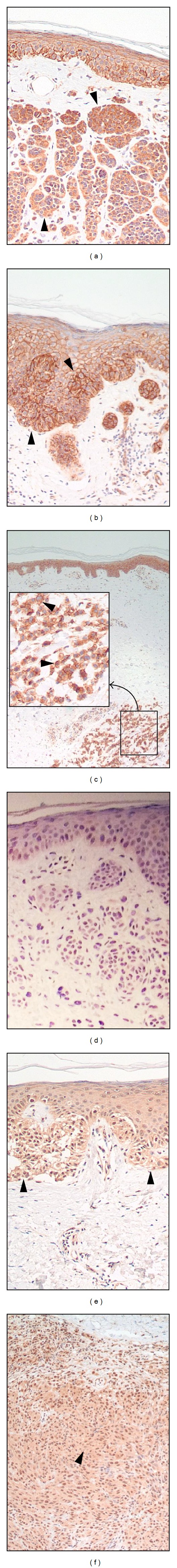
Representative pictures of β-catenin ((a)–(c)) and Rad6 ((d)–(f)) staining in nevus ((a), (d)), primary melanoma ((b), (e)), and metastatic melanoma ((c), (f)). Closed arrowheads point to positively immunostained cells. The highlighted square in panel (c) is magnified. Original magnification ×400.