
Figure 3.
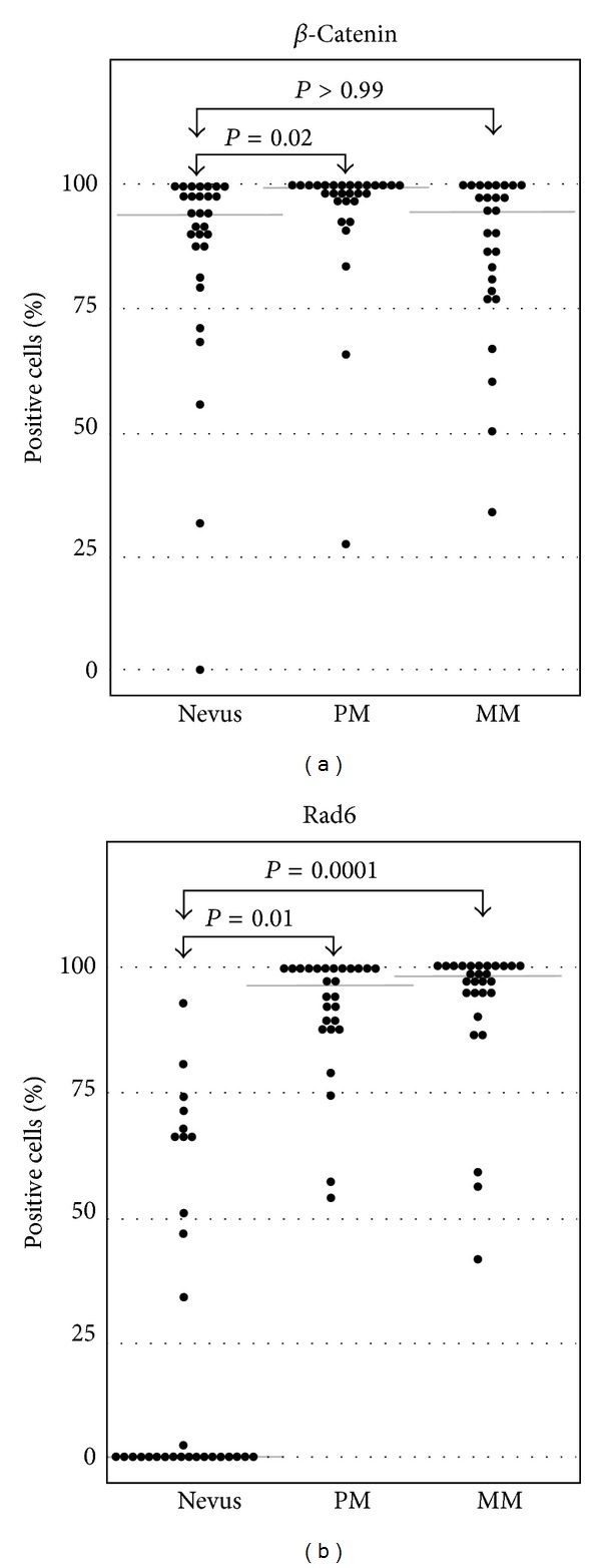
Boxplots of Rad6 and β-catenin positive cells in nevi, primary melanoma (PM), and metastatic melanoma (MM). Kruskal-Wallis tests showed that there are significantly more Rad6 positive cells in primary and metastatic melanomas as compared to nevi. Median values are indicated by gray horizontal lines.