
Figure 3.
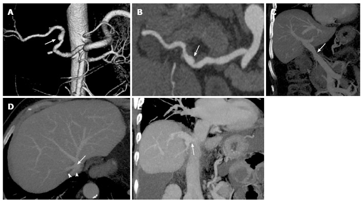
Common vascular anatomy after liver transplant on multidetector computed tomography images. Volume rendering reconstruction (A) and curved-maximum intensity projection (MIP) reconstruction (B) show conventional “fish-mouth” end-to-end anastomosis (arrow) between donor and recipient arterial vessels. Portal vein (C) is usually reconstructed with an end-to-end anastomosis (arrow) between donor and recipient portal veins, as shown in the coronal MIP reconstruction. “Piggyback” reconstruction (D) of the inferior vena cava (IVC), in which an end-to-side anastomosis is performed between the donor IVC (arrow) and the common stump of recipient hepatic veins (arrowheads). The anastomotic site is indicated by the arrow in the coronal MIP reconstruction (E).