Figure 5.

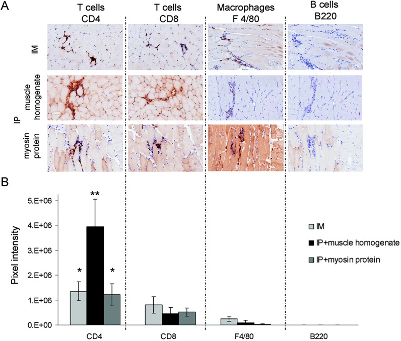
Inflammatory responses in myositis induced by exposure to muscle protein are predominated by CD4+ cells. Preparations of lymph node cells from FoxP3-deficient mice were adoptively transferred into recombination-activating gene 1–null recipients by intramuscular (IM) or intraperitoneal (IP) injection, with or without coadministration of muscle homogenate or purified myosin protein. A, Expression of CD4, CD8, F4/80, and B220 within inflammatory infiltrates, as determined by immunohistochemistry. Original magnification × 200. B, Results of digital image analysis showing the pixel intensity values in areas of infiltration for each leukocyte marker. Results are representative of similar trends observed in at least 5 mice (n = 4 independent experiments). Values are the mean ± SD. ∗ = P < 0.05; ∗∗ = P < 0.0001 versus all other markers in the same experimental group.
