
Figure 3.
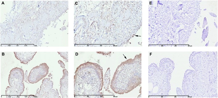
Immunohistochemical detection of hyaluronan synthase 1 (HAS-1) in normal/reactive (A and C) and inflamed (B and D) synovial biopsy samples. Also shown are negative controls with normal goat IgG in normal/reactive (E) and inflamed (F) synovial biopsy samples. In inflamed synovial biopsy samples, there is an increase in HAS-1–positive cells with predominant localization in the intima lining. At the time of surgery, synovial biopsy samples from normal/reactive or inflamed areas were macroscopically selected as described in Patients and Methods. Normal/reactive and inflamed synovial biopsy samples were stained with anti–HAS-1 antibody. Representative images are shown. Arrows indicate intima lining. Original magnification × 5 in A and B; × 10 in C–F.