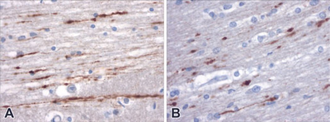
FIG. 3.
In two cases in which there was not inflicted cerebral trauma, multiple foci of labeled axons were demonstrated. Panel A demonstrates multiple longitudinal profiles of labeled axons within the temporal lobe of a child who was resuscitated and survived for almost a day after a near drowning (Case #15, 400× magnification). Panel B shows similar profiles of labeled axons within the white matter of sudden unexplained death in an infant who was resuscitated and survived for about 12 h (Case #12, 400× magnification).

