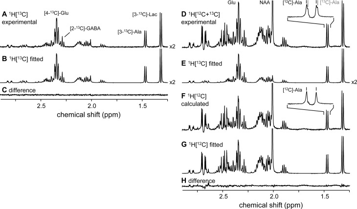
Figure 1.
Experimental (A) and fitted (B) 1H-[13C] NMR spectra from rat brain extract. (C) Difference (A–B) spectrum. (D) Experimental, total 1H-[12C+13C] NMR spectrum minus the fitted 1H-[13C] NMR spectrum (B/E) gives a calculated 1H-[12C] NMR spectrum (F), which can be fitted without a 13C isotope shift (G). (H) Difference (F–G) spectrum. The 13C isotope shift is visualized for alanine in panel D, but can be detected for all metabolites. The inset in panel F shows that the doublet signal from [3-13C]-alanine is effectively eliminated during the subtraction (D–E), leaving only the doublet signal from [3-12C]-alanine.

