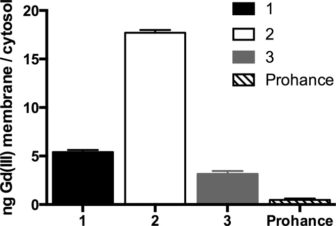
Figure 4.
Localization of contrast agents determined by cell fractionation. The internalized (cytosol + endosomes) and membrane fraction were analyzed by ICP-MS for Gd(III) content. All lipophilic complexes show higher membrane accumulation than Prohance with 2 having the greatest membrane localization. Error bars represent ± standard deviation of the mean of triplicate samples.

