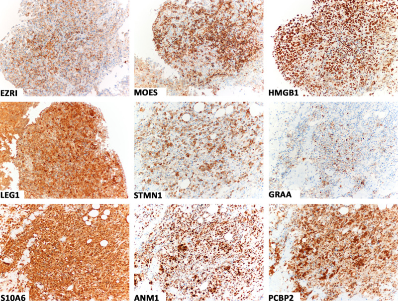
FIGURE 1.

Immunostains showing tumor cell positivity for ezrin (EZRI), moesin (MOES), high-mobility group box 1 (HMGB1), galectin 1 (LEG1), and stathmin 1 (STMN1) in case 3, and granzyme A (GRAA), S100 calcium-binding protein A6 (S10A6), protein arginine methyltransferases 1 (ANM1), and poly(rC)-binding protein 2 (PCBP2) in case 2. Almost all tumor cells were stained; the intensity of staining was usually strong (immunoperoxidase, hematoxylin counterstain).
