Figure 8.
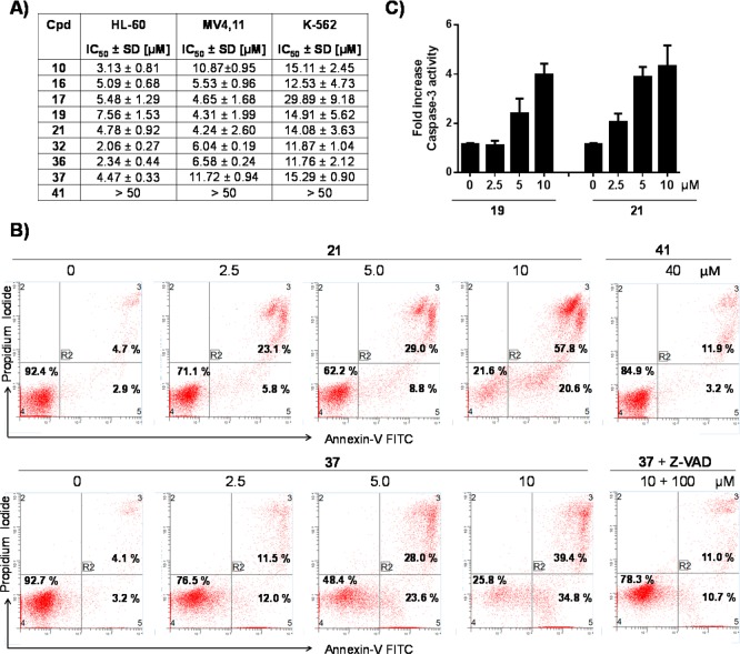
Cell death and apoptosis induction by Mcl-1 inhibitors in human leukemic cell lines. (A) Inhibition of cell growth by designed Mcl-1 inhibitors in the HL-60, MV4,11, and K-562 leukemia cell lines. Cells were treated for 3 days, and cell growth was determined using CellTiter Glo luminescent cell viability assay. (B) Analysis of apoptosis induced by 21 and 37 in the HL-60 leukemia cell line. Cells were treated with 21, 37, and 41 for 20 h using indicated concentrations, and apoptosis was analyzed with annexin-V and propidium iodide (PI) double staining by flow cytometry. Early apoptotic cells were defined as annexin-V positive/PI-negative, and late apoptotic cells as annexin-V/PI-double positive. Induction of the apoptosis by 37 was tested also in the presence of Z-VAD-FMK. (C) Induction of caspase-3 by 19 and 21 in the HL-60 cell line. Cells were treated for 20 h, and caspase-3 was detected with fluorometric-based assay. Results shown are the mean and SEM from at least three separate experiments.

