
Figure 2.
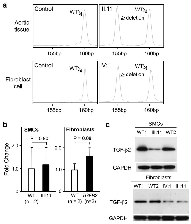
Transcript and protein analysis of the TGFB2 mutation p.Tyr341Cysfs*25 in exon 6. (a) RT-PCR of RNA extracted from normal ascending aortic specimens (n=2) or fibroblast cell cultures (n=2) showed only one PCR product at 160 bp. RT-PCR of RNA extracted from ascending aortic specimen of a patient (III:11) or fibroblast cell culture (IV:1) from family TAA288 demonstrated that the transcript from both normal and 5 bp deleted allele were present. (b) Expression of TGFB2 was quantified in aortic SMCs and dermal fibroblasts explanted from TGFB2 mutation carriers and matched controls using quantitative-PCR. TGFB2 expression levels between patients and controls were similar in both SMCs and fibroblasts. TGFB2 levels are standardized to GAPDH messages. The relative expression values were determined via the ΔΔCt method, and assays were performed in triplicate. Data are expressed as mean ± standard error of the mean for pooled experimental results. (c) Immunoblot analysis of TGF-β2 proprotein in cellular lysates of aortic SMCs and fibroblasts from patients (III:11 and IV:1, TAA288 family) and two normal controls. A protein band at 47kD, the molecular weight of the proprotein of TGF-β2, was identified with immunoblot analysis in both the patient and control lysates using a polyclonal antibody specific for TGF-β2. Decreased intensity of this band was found in patients’ cells, both SMCs and fibroblasts, when compared with control cells.