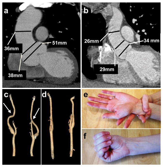
Figure 4.
Clinical features associated with TGFB2 mutations. (a) CT scan imaging of the aorta of patient III:16 from family MS239 demonstrates dilatation predominating at the level of the sinuses of Valsalva (50 mm). (b) CT scan of a normal aorta with a diameter of 34 mm at the level of the sinuses of Valsalva. (c) Three dimensional CT scan from patient MS239, III:16 showing mild tortuosity (arrows) of cerebral arteries compared to normal control (d). Minimal arachnodactyly is evident in individual TAA288 III:8 based on positive wrist (Walker) sign (e) but negative thumb (Steinberg) sign (f).

