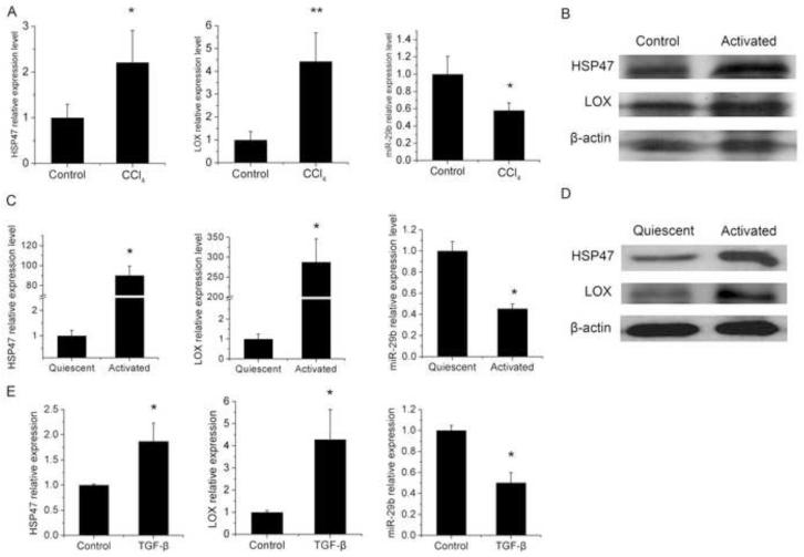
Fig. 1. Expression of HSP47, LOX and miR-29b in mouse liver with CCl4-induced fibrosis, culture-activated rat HSCs and TGF-β treated LX-2 cells.
CD-1 mice were treated with corn oil or CCl4 for 6 weeks (A-B). Quiescent and activated HSCs of rat were isolated and harvested as described in the Materials and methods (C-D). LX-2 cells were treated with TGF-β (5ng/ml) and harvested at 24h after treatment (E). Quantitative PCR was conducted to detect the expression levels of HSP47 and LOX mRNA and miR-29b. Gene expression level was normalized against the control groups, and data represent quantification of four independent experiments, *P < 0.05 (A, C and E). Western blots were conducted to detect the protein expression levels of HSP47 and LOX (B and D).

