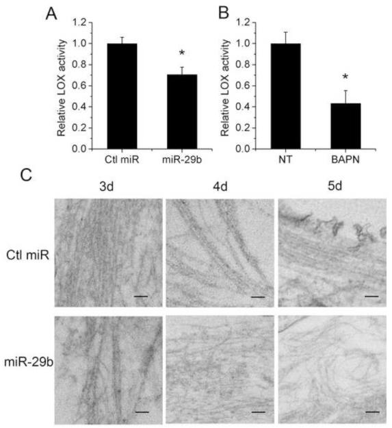
Fig. 3. Transfection of LX-2 cells with miR-29b affects their extracellular LOX activity and morphology of extracellular fibrils.
(A) Extracellular LOX enzyme activity was significantly reduced in the supernatant of miR-29b transfected LX-2 cells 72 h post-transfection compared with that of control miRNA transfected cells. (B) LOX activity in the conditioned media was significantly inhibited by 100 μM BAPN. Data represent mean ± SD, n = 3. (*P < 0.05). (C) LX-2 cells were transfected with control miRNA or miR-29b. The extracellular fibrils were observed by Transmission Electron Microscopy at 3 d, 4 d and 5 d after the cells became confluent. Bar, 100 nm.

