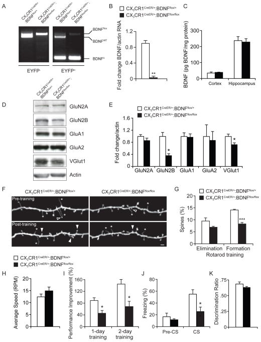
Figure 5. Loss of microglial BDNF results in altered synaptic protein levels, synaptic structural plasticity, and performance improvement after learning.
(A) PCR-based analysis of wild-type (BDNFWT), conditional undeleted (BDNFflox), or conditional deleted (BDNFΔ) BDNF alleles from CX3CR1-EYFP− and CX3CR1-EYFP+ cells sorted from the CNS of CX3CR1CreER/+:BDNFflox/+ or CX3CR1CreER/+:BDNFflox/flox after tamoxifen treatment. (B) Quantitative real-time PCR analysis of BDNF mRNA isolated from CX3CR1-EYFP+ microglia purified from BDNFflox/+ or BDNFflox/flox mice (**p<0.01, n=3). (C) Average protein levels of total BDNF in the cortex or hippocampus of CX3CR1CreER/+:BDNFflox/+ or CX3CR1CreER/+:BDNFflox/flox mice as measured by ELISA (n=4). (D) Synaptosome fractions from the brains of CX3CR1CreER/+:BDNFflox/+ or CX3CR1CreER/+:BDNFflox/flox mice probed with indicated antibodies. (E) Densitometric quantification of western blots in (D) (*p<0.05, n=6). (F) Transcranial two-photon imaging of dendritic spines in Thy1 YFP mice crossed with CX3CR1CreER/+:BDNFflox/+ or CX3CR1CreER/+:BDNFflox/flox mice before or after rotarod training. Filled and empty arrowheads indicate spines formed or eliminated between two views. Asterisk indicates filopodia. Scale bar= 2 μm. (G) Percentage of existing spines eliminated, or new spines formed over 2 days of training in the motor cortex of BDNFflox/+ or BDNFflox/flox mice (***p<0.001, n=4). (H) Average speed reached during the first rotarod training session (n=5–7). (I) Performance increase in motor learning task over 1 or 2 days of rotarod training (*p<0.05, error bars= SEM, n=5–7). (J) Percentage of freezing in control CX3CR1CreER/+:BDNFflox/+ or CX3CR1CreER/+:BDNFflox/flox mice before (pre-CS) and during (CS) presentation of the conditioned stimulus in the recall test (*p<0.05, n=6–7) (K) Discrimination ratio of time spent interacting with a novel object vs. a familiar object in a novel object recognition assay was significantly altered in mice depleted of microglial BDNF (*p<0.05, n=8). Data are represented as mean +/− SEM. See also Figure S5.

