
Figure 2.
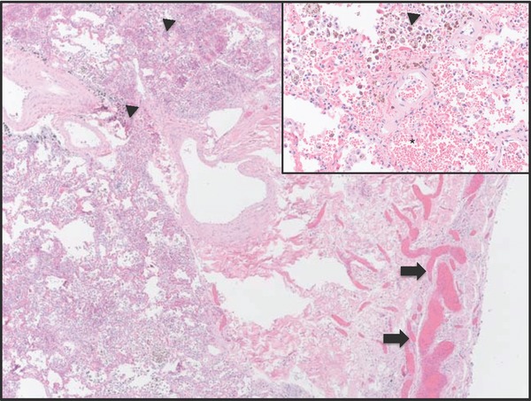
Histology of lung (hematoxylin and eosin, 12.5×). There are very dilated bronchial veins (arrows) and hemosiderin‐laden macrophages (arrowheads). Inset: High power (400×) view of hemosiderin‐laden macrophages and intra‐alveolar hemorrhage (*).