
Figure 4.
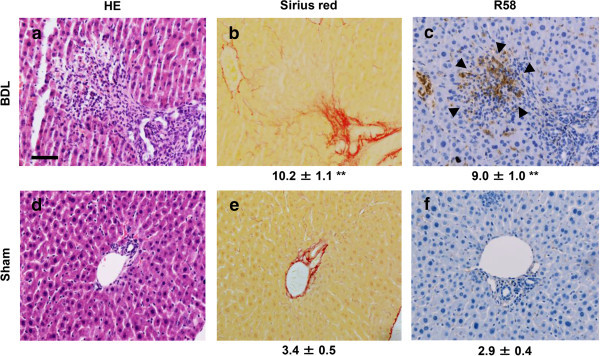
Immunohistochemistry of R 58 LAP-DP in fibrotic liver tissues in BDL mice using R58 antibody. Liver tissues from BDL and sham-operated mice were stained by HE (a, d) and Sirius Red (b, e), and then immunostained with R58 antibody (c, f). Scale bar = 50 μm. The percentage of fibrotic regions and R58 positive areas were calculated from three fields each from five sections (5 mice) (total 15 fields) and described as average ± SE under the corresponding panels. A total of 16 mice were evaluated and representative results are shown.