
Figure 6.
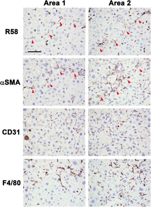
Identification of R58 positive cells in CCl 4 -treated mice liver sections. Paraffin serial sections were immunostained with, R58 (upper panels), anti-αSMA (second row panels), anti-CD31 (third row panels), and anti-F4/80 (lower panels) antibodies. R58 positive signals were compared with αSMA, CD31, or F4/80 signals in same areas of each sections. The red arrowheads (R58 and αSMA panels) represent respectively both R58 and αSMA positive cells along with fibrotic septa. Scale bar = 50 μm.