
Figure 7.
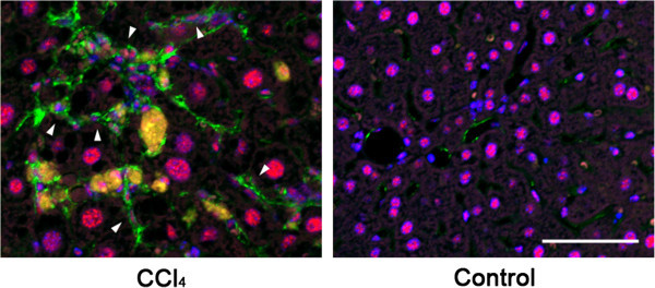
Detection of phosphorylated Smad3C in activated HSCs in CCl 4 -treated mice liver sections. Phosphorylation and nuclear translocation of Smad3C in activated HSCs were examined by immunofluorescent double staining with anti-pSmad3C (red) and anti-αSMA (green) antibodies. Nuclei of the cells were identified by Hoechst 33258 staining (blue). The pSmad3C positive-activated HSCs were seen only in CCl4-treated mice. Scale bar = 50 μm.