
Figure 8.
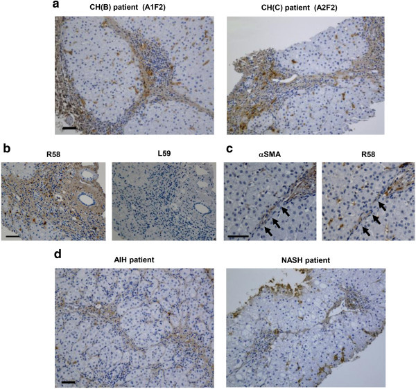
Immunostaining of biopsied human liver tissues. (a, d) Liver biopsy samples from four patients (chronic hepatitis B and C virus infection [CH (B) and (C), respectively], AIH with severe fibrosis, and NASH. The hepatitis activity grade and fibrosis score are described in the upper corresponding micrograms. (b) Serial sections from CH(C) patient were stained with R58 and L59 antibodies. (c) Liver specimens from another CH(B) patient were stained with anti-αSMA, R58 antibodies. αSMA and R58 signal positive cells lined up along fibrous septa (yellow arrows). Scale bars = 50 μm.