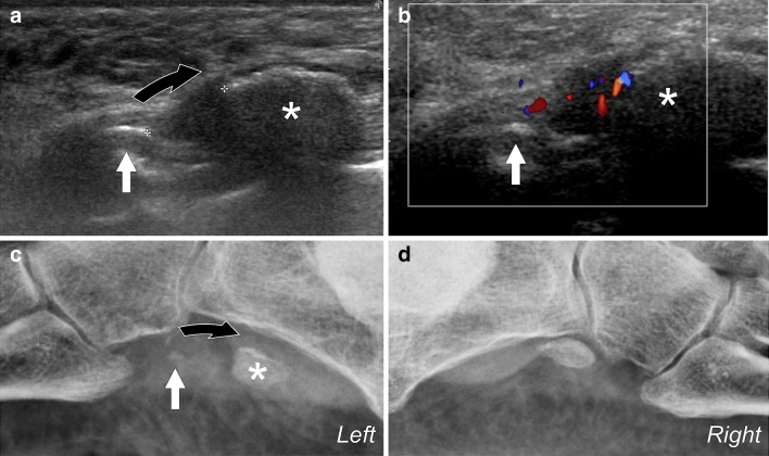
Fig. 13.
a Longitudinal US scan obtained at the os peroneum in a patient with type 2 tendon tear. The proximal bone fragment (big asterisk) is larger than the distal fragment (white arrow) and is proximally dislocated (curved arrow) due to the traction exerted by the peroneus longus tendon. The dislocation can be evaluated at US imaging (calipers). The distal fragment, which is smaller, seems not to be dislocated. b Color Doppler shows significant hyperemia present in the fracture. c Plain film X-ray confirms US findings showing dislocation of the bone fragments. d Plain film X-ray of the contralateral normal ankle. Note the correct position of the os peroneum at the calcaneal cuboid joint

