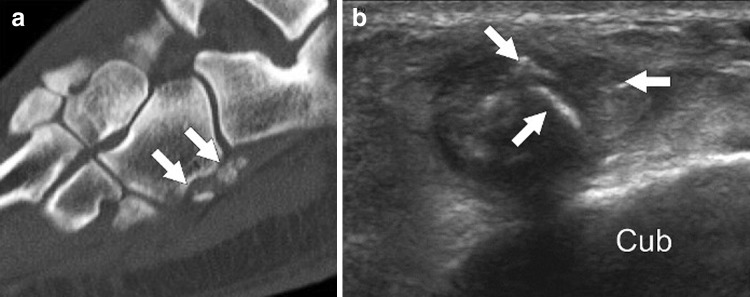
Fig. 9.
Multipartite os peroneum. Computed tomography (sagittal reconstruction) (a) and US coronal oblique scan (b) in a patient with multipartite os peroneum (arrows). The tubercle presents a bifid appearance (asterisks). Note the characteristic sclerotic, quite sharp margins of the bone fragments which are located close to each other near the base of the fifth metatarsal bone. Cub cuboid

