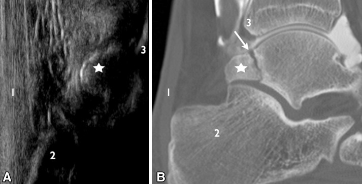
Fig. 1.
Posterior ankle impingement. a Longitudinal scan showing the posterolateral process of the talus (star) with no sign of synovitis. b CT arthrography with reconstruction in the sagittal plane showing rupture due to pseudoarthrosis (white arrow) in the posterolateral process (star), not identified on US image. 1 Achilles tendon; 2 calcaneus; 3 tibia

