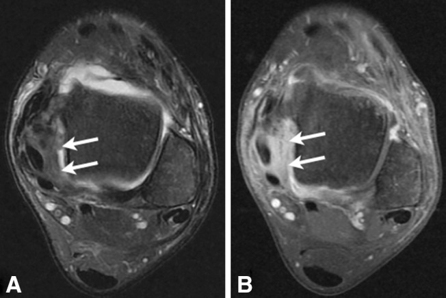
Fig. 3.

Posteromedial ankle impingement. MR imaging, axial scan, T2-weighted with fat saturation (a) and T1-weighted with fat saturation after gadolinium injection (b), shows the presence of inflammatory synovitis (white arrows) on the posterior aspect of the medial capsular ligaments
