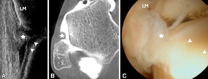
Fig. 5.

Anterolateral ankle impingement. US imaging, axial-oblique view (a), CT arthrography, oblique axial reconstruction (b) and arthroscopy (c) show the presence of nodular synovitis (star) adjacent to the cartilaginous portion of the superolateral aspect of the talus (arrowheads). LM lateral malleolus
