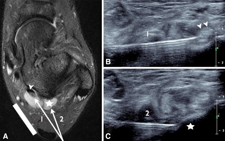
Fig. 9.
Anterolateral ankle impingement, injection of anesthetics. a Axial T2-weighted MR scan with fat saturation shows synovitis of the posterior recess of the ankle (star) and fluid collection in the sheath of the flexor hallucis longus tendon (arrowhead). US imaging: probe position (white line), needle path under US guidance and axial scans to monitor the needle at the flexor hallucis longus tendon (b) and the posterolateral process of the talus (c)

