
Figure 1.
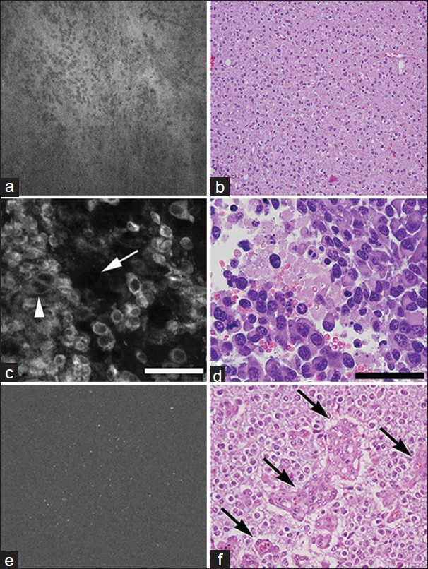
Images obtained with intraoperative endomicroscopes of various clinically available fluorescent contrast agents. (a and b) Fluorescein-induced fluorescence of oligodendroglioma (Grade II), and corresponding H and E stain. (c and d) ICG-induced fluorescence of glioblastoma cells in a mouse model, and corresponding H and E stain. (e and f) 5-ALA induced fluorescence in low-grade glioma, and corresponding H and E stain. Figures a and b from Eschbacher et al.;[24] used with permission from Journal of Neurosurgery. Figure c used with permission from Barrow Neurological Institute. Figure d from Martirosyan et al.;[64] used with permission from Journal of Neurosurgery. Figures e and f from Sanai et al.;[87] used with permission from Journal of Neurosurgery