Figure 1.
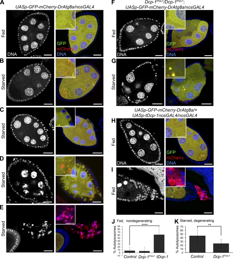
Dcp-1 is necessary for autophagic flux during midoogenesis. GFP-mCherry-DrAtg8a was expressed in the germline using the nosGAL4 driver. Staining shows DNA, GFP, and mCherry. (A) UASp-GFP-mCherry-DrAtg8a/+;nosGAL4/+ flies conditioned on yeast paste had diffuse GFP-mCherry-DrAtg8a staining in midstage egg chambers. (B) Nondegenerating midstage egg chambers from starved flies contained autophagosomes (yellow) and autolysosomes (red). (C and D) Egg chambers early in the degeneration process showed follicle cells that take up portions of the nurse cell cytoplasm (C) followed by condensation and fragmentation of the nurse cell nuclei and further uptake of the nurse cell cytoplasm into follicle cells (D). (E) Late stage degenerating egg chambers lose all GFP staining and fluoresce red. (F) Fed Dcp-1Prev1/Dcp-1Prev1;UASp-GFP-mCherry-DrAtg8a/nosGAL4 flies showed diffuse GFP-mCherry-DrAtg8a staining in the germline. (G) Starved Dcp-1Prev1/Dcp-1Prev1;UASp-GFP-mCherry-DrAtg8a/nosGAL4 flies showed reduced autolysosomes in degenerating midstage egg chambers. (H and I) Fed flies overexpressing truncated Dcp-1 (tDcp-1) in the germline showed increased autophagosomes and autolysosomes in nondegenerating midstage egg chambers (H) and also contained degenerating midstage egg chambers that lose all GFP fluorescence and fluoresce red (I). Bars: (main images) 25 µm; (insets) 12.5 µm. Insets in A–I show diffuse cytoplasmic staining of Atg8a or autophagosomes and autolysosomes. (J and K) The percentages of autolysosomes (autolysosomes/total autophagic structures) were manually calculated in at least eight egg chambers for each genotype as indicated. Error bars represent the means ± SD. Statistical testing was performed using one-way ANOVA with a Dunnet post test (****, P < 0.0001) or a two-tailed Student’s t test (**, P < 0.005).

