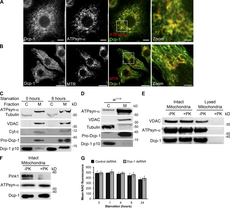
Figure 4.
Dcp-1 is partially localized within mitochondria. (A and B) l(2)mbn cells were labeled with antibodies to Dcp-1 and ATPsyn-α (A) or MitoTracker red (MTR; B). Merged images show colocalization between Dcp-1 and the mitochondria. Boxes represent zoomed images. Bars: (main images) 5 µm; (zoomed images) 1.25 µm. (C) Western blot from l(2)mbn cells subjected to nutrient-rich or starvation conditions for 6 h. Cells were separated into cytosolic (C) and mitochondrial enriched (M) fractions. (D) Ovaries from fed w1118 flies were separated into cytosolic and membrane-enriched fractions and probed with antibodies to VDAC, Tubulin, ATPsyn-α, and Dcp-1. (E and F) Intact and lysed mitochondria (E) or intact mitochondria isolated from l(2)mbn cells (F) were treated with proteinase K (PK). The effects of proteinase K treatment were assessed by antibodies to VDAC, ATPsyn-α, Pink1, and Dcp-1. (G) Control and Dcp-1 RNAi–treated cells were subjected to nutrient-rich or starvation conditions and stained with NAO. Mean fluorescence was measured by flow cytometry. Graph represents ± SEM of three independent experiments (n = 3).

