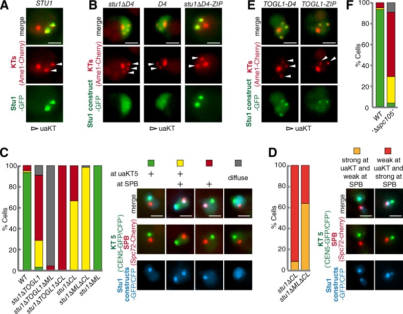
Figure 3.
Stu1 localization to uaKTs depends on TOGL1, CL, dimerization, and Spc105. (A–F) Cells were analyzed 3 h after release from G1 into medium containing Nz. Bars, 2 µm. (A) Stu1 selectively associates with uaKTs. (B) Dimerization is required for Stu1 localization to uaKTs. To analyze D4-GFP cells, WT Stu1 expressed in the background was depleted. (C and D) TOGL1 and CL are essential for Stu1 localization to uaKTs. (C) Phenotypes were quantified as indicated in cells with a uaKT5. n > 70. (D) More detailed analyses of the Stu1 signal intensity at SPBs and uaKTs. n > 70. (E) TOGL1 interacts with uaKTs. WT Stu1 expressed in the background was depleted. (F) uaKT localization depends on Spc105. Depletion of Spc105 (‘Δspc105’) was as described in the Materials and methods section. Phenotypes were quantified as in C. n = 72. The result obtained for WT cells is shown as a comparison.

