
Fig. 1.
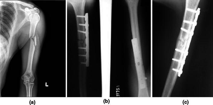
a Preoperative X-ray of 30-year-old male patient showing displaced fracture of the shaft of the humerus, left side. b Immediate postoperative X-ray anteroposterior (AP) and lateral view showing plate osteosynthesis with 4.5-mm narrow LCDCP. c Postoperative X-ray at 12-week follow-up visit showing a well-uniting fracture