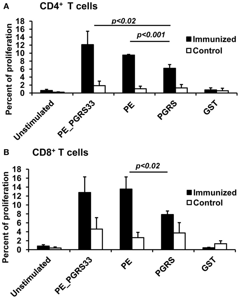
Figure 2.
Antigen-specific proliferation of CD4+ and CD8+ T cells in immunized mice. Splenocytes from PE_PGRS33-immunized mice were stained with CFSE and incubated with 25 μg of PE_PGRS33, PGRS, PE, or GST protein plus 10 μg of Polymyxin B for 4 days. Spleen cells from mice injected with only nitrocellulose were also cultured with antigens (control). Cells without the antigens were incubated for the same length of time (unstimulated). Splenocytes were then labeled with anti-CD4-phycoerythrin (A) or anti-CD8-allophycocyanin (B) monoclonal antibodies and the percentage of proliferating cells were determined by CFSE dilution and flow cytometry. Each bar represents the mean ± SD of data from four mice per group, and the results are representative of those obtained from three independent experiments.

