
Figure 1.
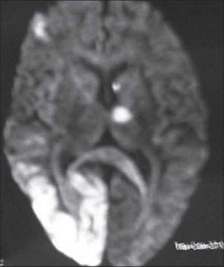
Magnetic resonance imaging of the brain showing acute ischemic infarcts in left cerebellar hemisphere, bilateral occipital lobe and left thalamus
Official websites use .gov
A
.gov website belongs to an official
government organization in the United States.
Secure .gov websites use HTTPS
A lock (
) or https:// means you've safely
connected to the .gov website. Share sensitive
information only on official, secure websites.
Magnetic resonance imaging of the brain showing acute ischemic infarcts in left cerebellar hemisphere, bilateral occipital lobe and left thalamus