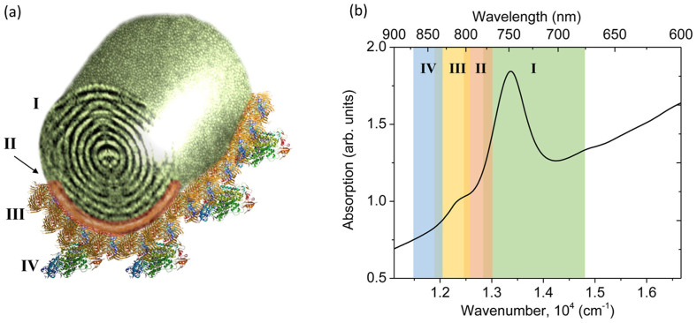
Figure 1.
(a) Schematic structure of the light-harvesting complex (LHC) in green sulfur bacteria: I –chlorosome; II – baseplate; III – Fenna-Matthews-Olson protein complexes; IV – reaction centers. (b) Qy-band absorption of a bacterial culture, the marked ranges correspond to different structural units of LHC. In Cba. tepidum the structural unit I is composed of BChl c pigments, while the units II–IV contain BChl a pigments.

