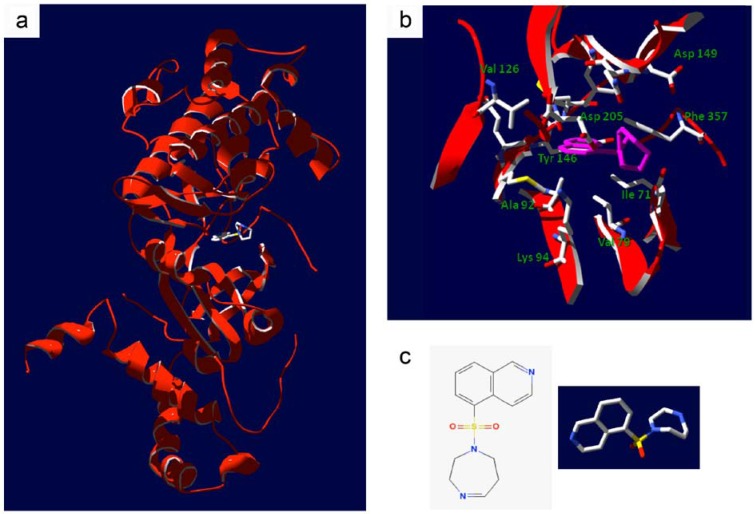
Figure 3.
Structure of the ROCK kinase domain in complex with fasudil. (a), Ribbon diagram detailing the crystal structure of the bovine ROCK kinase domain in complex with fasudil, (residues 18 to 417, PDB ID: 2F2U [34]). (b), Detail of fasudil occupancy within the ATP binding pocket of ROCK; amino acid side chains lining the pocket within 6 Å of fasudil are shown. Fasudil is shown in pink. (c), The molecular structure of fasudil in 2- and 3-dimensional renderings.

