
Figure 3.
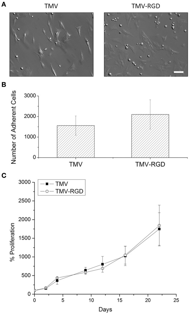
BMSC adhesion and viability on virus scaffolds. (A) Optical images of BMSC attached on virus substrates after 24-h seeding in primary media. Scale bar is 100 μm. (B) Number of adherent BMSCs on different virus substrates after 24-h seeding in primary media. (C) Proliferation percentage of BMSCs on TMV and TMV-RGD substrates over 22 days in osteogenic media. Error bars indicate ±1 SD.