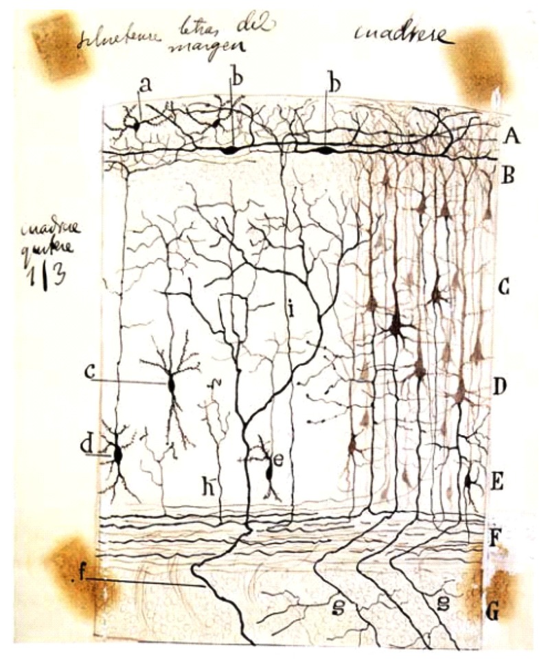
FIGURE 1.
Schematic drawing by Cajal of a Golgi-impregnated preparation of the cerebral cortex. In this illustration, Cajal compiled some of his findings from small mammals (rabbit, mouse, etc.) reported between 1890 and 1891. Note both the presence of the polyhedral (or stellate) cells (a) and the horizontal fusiform cells (b) in the plexiform layer. A, plexiform layer; B, small pyramidal cell layer; C, medium pyramidal cell layer; D, giant pyramidal cell layer; E, polymorphic cell layer; F, white matter; G, striatum. Reproduced, with permission of the Inheritors of Santiago Ramón y Cajal, from Reference (Ramón y Cajal, 1923).

