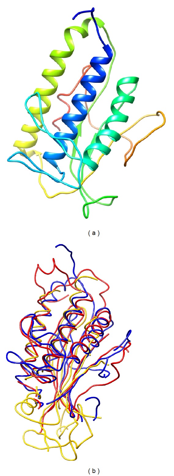
Figure 8.

Ribbon representation of the best model of Drosopohila Atg101 obtained with I-TASSER (a). Atg101 structure alignment of the best model obtained with I-TASSER (blue), as well as human Mad2 (2V64, chain A, yellow) and Atg13 from the yeast Lachancea thermotolerans (4J2G, chain A, red) (b).
