
Fig. 1.
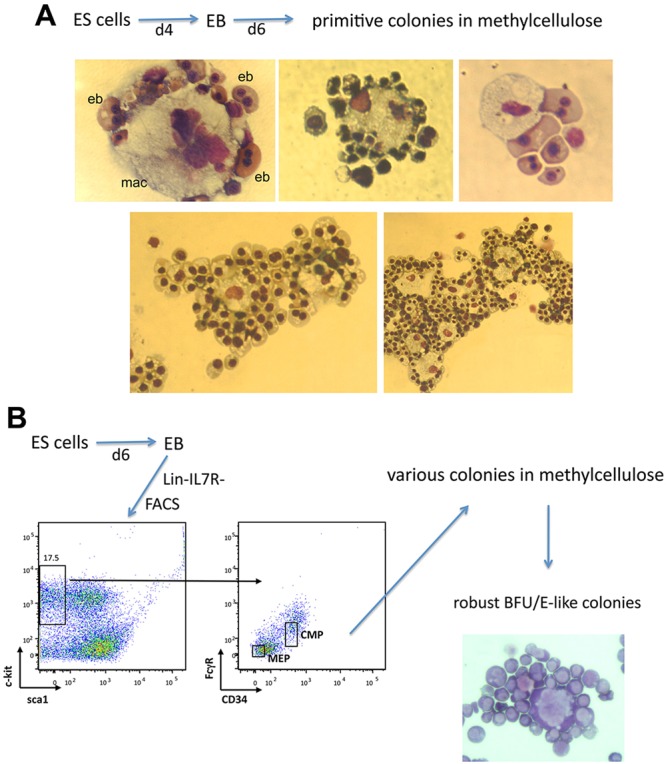
Typical morphology of erythroid colonies derived from differentiating ESCs. Individual methylcellulose erythroid colonies from (A) secondary platings of dispersed EBs or from (B) FACS-sorted dispersed EBs (enriched for MEP or CMP populations as shown) were picked and cytospun onto a glass slide. The slides were air dried and stained with May-Grünwald Giemsa. Note that not all contain a single island (A, bottom); some EB-derived erythroid colonies contain two (left) or more (right) central macrophage. eb, erythoblast; mac, macrophage.