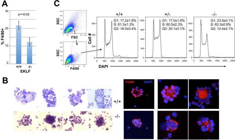
Fig. 6.
Erythroblastic island integrity is dependent on EKLF. Tests used cells derived from wild-type (+/+), EKLF-het (+/−) or EKLF-null (−/−) E13.5 fetal livers as indicated. (A) F4/80+ macrophage cellularity was monitored by FACS and displayed as percentage of total fetal liver cells. (B) Visual analysis of isolated erythroblastic islands (+/+ top row; −/− bottom row). Left, May-Grünwald Giemsa stain of four typical islands from each. Right, macrophage F4/80 stain (red) and DAPI DNA stain (blue) from three typical islands from each (two different magnifications are shown). (C) Cell cycle analysis of dispersed cells is shown.

