
Fig. 7.
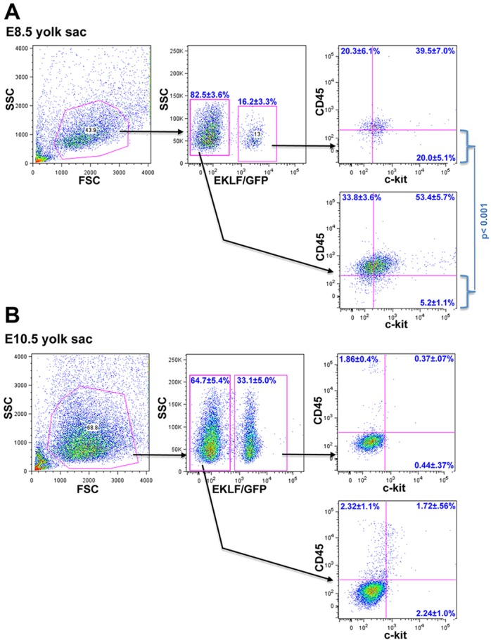
Analysis of EMP and progeny in sorted EKLF/GFP+ or EKLF/GFP− yolk sac cells in early development. Yolk sacs from E8.5 (A; n=5) or E10.5 (B; n=5) embryos were dispersed and analyzed for EKLF/GFP, CD45 and Kit (CD117) expression by FACS. The statistically significant different percentages of CD45–/Kit+ levels between EKLF/GFP+ and EKLF/GFP− cells (i.e. lower right quadrant in each case) are shown for E8.5 (A).