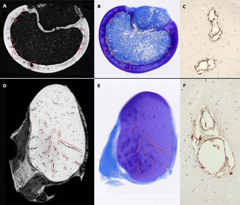
Figure 1.
Ex vivo SWI images (Panels A and D) obtained using a 9.4 T MRI scanner and corresponding toluidine blue stained histological slides (panels B and E) of the sagittal ridge of the humeral trochlea of a 6-week-old pig (upper panels, sectioned in the sagittal plane) and medial femoral condyle of a 3-week-old pig (lower panels, sectioned in the transverse plane). Red markers identify corresponding cartilage canal vessels. Immunostaining of adjacent sections using an antibody directed against Von Willebrand Factor (Panels C and F; imaged with a 20× objective) demonstrated positive immunostaining (brown reaction product) in endothelial cells lining cartilage canal vessels.

