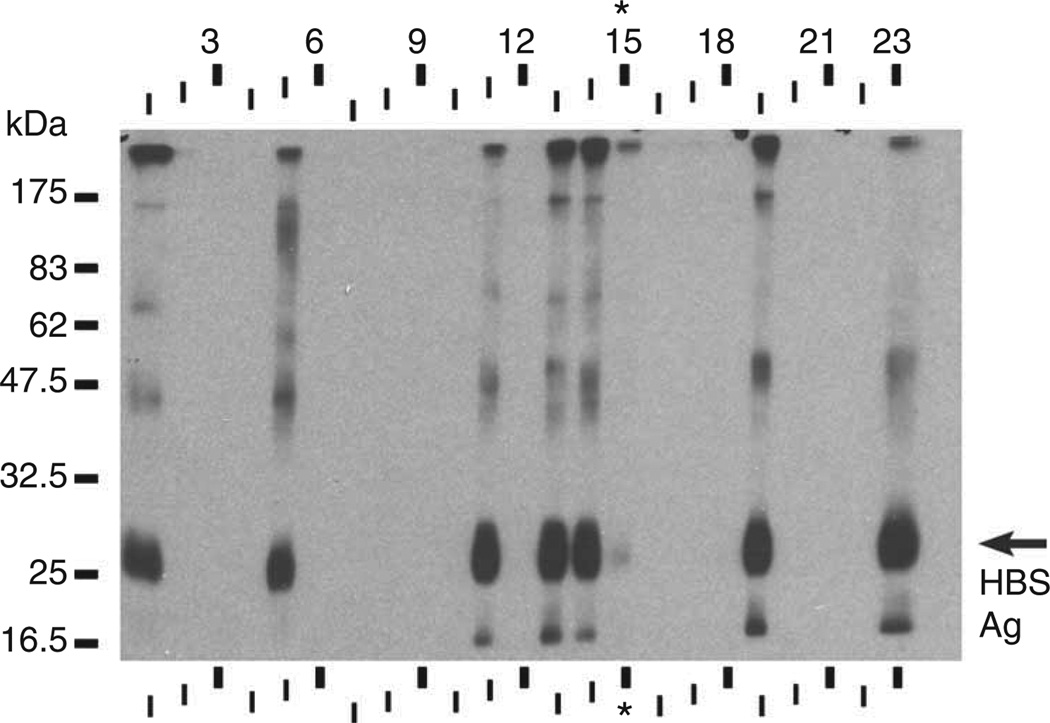
Figure 5.
‘Curtain’ western blot of 12.5% polyacrylamide gel loaded with 200 µg of complete COS-1 cell lysate with 10 µg HBSAg (ViroStat). Using a Surfblot device (Idea Scientific), a different culture supernatant was applied to each strip, numbered 1–23, except lane 15 to which sera from the immunized mouse (1:200 dilution) was applied (asterisk). The film for the blot shown was exposed for 60 s. A duplicate blot without added HBSAg was blank (data not shown).

